
Locations and Care
![]()
Whether you’re home or away, find a location closest to you. Search by facility.
Logan Services
Logan Health offers a wide variety of services and care. Please select from the dropdown below to see more specific to your health care needs.
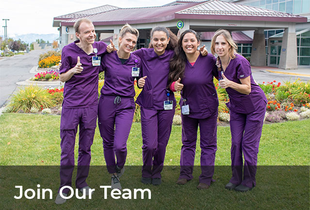
Our people are everything. The amazing providers, employees, and volunteers at Logan Health gift this organization with their innovation, knowledge, and compassion. Together, we’re connecting our community to trusted, quality care and helping to improve lives every day.

By offering high-quality health care, responding to community needs, and concentrating resources in areas that truly make a difference, Logan Health maintains a rich tradition of giving back to the communities we serve.
Logan News & Events
![]()
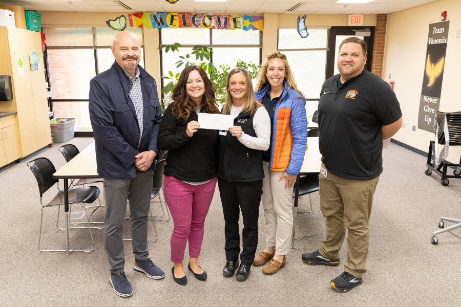
Logan Health awards Healthy Classrooms grants to 19 Flathead Valley educators
Logan Health’s Foundation and School-Based Services awarded $500 grants to 19 educators at Flathead Valley schools this April. The Healthy Classrooms Grant provides funding to local schools to aid in the creation or improvement of student-centered and staff-related healthy living initiatives.
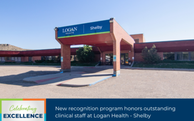
New recognition program honors outstanding clinical staff at Logan Health – Shelby
Logan Health – Shelby is excited to announce nominations for the Sunshine Award are now open! Patients, visitors and employees can nominate any Logan Health – Shelby (LHS) clinical staff, including non-nursing caregivers, based on how they enrich a positive atmosphere throughout the Logan Health community, demonstrating consistent performance above and beyond their daily workload, and always putting the patient first.

Breaking the Silence: Let’s Talk About It campaign aims to tackle senior suicide and mental health stigma
In Montana, suicide rates among seniors rank among the nation’s highest. Because of this, Billings Clinic-Logan Health is spearheading an initiative called the “Let’s Talk About It” campaign, which aims to not only address the pressing issue of suicide among the population, but also dismantle the stigma that surrounds mental health.
Community Impact: Health and Safety Education
By offering high-quality health care, responding to community needs, and concentrating resources in areas that truly make a difference, Logan Health maintains a rich tradition of giving back to the communities we serve.